Toward Nano-Scale Scanning Electrochemical Microscopy of Single Live Cells by Means of the COMSOL Multiphysics® software
Scanning Electrochemical Microscopy (SECM) is a powerful noninvasive analytical technique employed in a variety of applications ranging from electrochemical characterization of material surfaces, to bioanalytical characterization of single live cells [1]. When applied to live cells, SECM has shown great strength as a technique to study cell topography [2], membrane permeability [3] and extracellular reactive oxygen species (ROS) [4] which play very important roles many cell biological processes. SECM can provide great insight into the physiological characteristics of single live cells. The use of smaller electrodes, such as nano-scale electrodes, provides higher experimental resolutions. However, this also necessitates greater considerations when creating models to quantify the above characteristics. SECM is commonly coupled with simulations in the COMSOL Multiphysics® software, and models to perform these calculations have been actively developed in our research lab at The University of Western Ontario.
Redox species diffusion to a SECM ultramicroelectrode (UME) probe and consumption at the UME are handled by the Chemical Reaction Engineering module. The cell membrane regulated transport of this redox species is defined by a limited flux between the inner and outer solution domains. The integration of the species flux at the UME tip allows for the calculation of the UME tip current, which is plotted vs. cell to UME distance (probe approach curves (PACs)) or vs. UME plane coordinates (mapping). While fitting of simulated PACs to the experimental ones gives ROS concentrations and permeability, mapping provides excellent visualization of cell topography and activity.
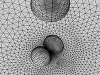
Our development of SECM live cell models has included tailored 3D sample geometry, allowing for the characterization of more complex sample features, such as nonuniform cell topography or the deconvolution of adjacent cell signals. Simulation of the full 3D mapping of a cell sample can be realized by parametric sweeping of the model geometry in COMSOL®. Our 3D models allow for the more accurate reconstruction of the cell membrane geometry and provide single cell maps of membrane permeability, and extracellular ROS concentrations. The above developments with COMSOL® simulations are versatile, and further strengthen SECM as an excellent bioanalytical tool. In this presentation, we demonstrate our very recent and exciting investigations on nano-scale SECM of single live cells by means of COMSOL®. We will provide guidelines on the research and development in this exciting field.
[1] F. P. Filice, M. S. M. Li, J. D. Henderson, Z. F. Ding, J. Phys. Chem. C 2015, 119, 21473. [2] M. S. M. Li, F. P. Filice, Z. F. Ding, J. Electroanal. Chem. 2016, 779, 176. [3] J. D. Henderson, F. P. Filice, M. S. M. Li, Z. F. Ding, ChemElectroChem 2017, 4, 856. [4] X. Zhao, P. M. Diakowski, Z. Ding, Anal. Chem. 2010, 82, 8371-8373.
下载
- filice_presentation.pdf - 10.24MB
- filice_abstract.pdf - 0.04MB