Simulating Small Molecule Transport Across the Blood-Brain Barrier Using COMSOL Multiphysics®
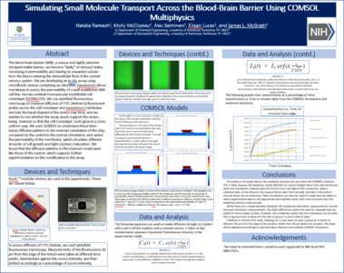
The blood-brain barrier presents a unique and highly selective semipermeable barrier composed of endothelial and supporting cells. In stressed states, such as during systemic inflammation, the barrier can become “leaky”, increasing in permeability and leading to unwanted solutes from the blood entering the extracellular fluid of the central nervous system where neurons reside. We are developing an assay using microfluidic devices containing an ultra-thin, nanoporous silicon membrane to assess the permeability of a human cerebral microvascular endothelial cell monolayer, which simulates the blood-brain barrier. These membranes are ideal for co-culture studies and in situ analyses requiring excellent optical properties. We use widefield fluorescence microscopy or confocal microscopy to measure diffusion of FITC-Dextran (a fluorescent probe) across the cell monolayer and nanoporous membrane and into the basal channel of the device over time. Traditional device orientation contains a “trench” facing up, which allows measurements in an open channel, but causes the cell monolayer to experience changes in geometry and grow in a non-uniform way.
To alleviate this mechanical stimuli, we are growing cells on the flat side of the chip, with the trench facing down. This orientation allows growth of a flat monolayer that more closely resembles the blood-brain barrier; however, it poses a more complex pattern of diffusion within the trench, where we are taking our dye measurements, compared to within the channel. To understand these diffusion patterns, we used COMSOL®, to take a 2D slice of the device in the trench down orientation and calculated the time-based transport of FITC-Dextran, while also varying the permeability of the membrane, which simulates different amounts of cell growth and tight junction maturation. We compared this to a control simulation of the device in the traditional orientation. We found that the diffusion patterns in the reversed model were similar to those of the control, which supports further experimentation on this modification to the assay.