Trapping of Single-Cells Within 3D Electrokinetic Cages
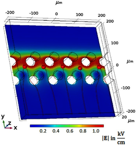
This paper reports the modelling and analysis of three dimensional negative dielectrophoretic traps for cell trapping applications. Dielectrophoresis is a well-established technique for cell analysis and cell trapping. Planar electrodes, at the bottom of a microfluidic channel, have been used for many years. New fabrication methods allow to produce three-dimensional electrodes which can be freestanding or integrated in the wall of a microfluidic channel. These electrodes provide homogeneous electric fields and therefore a uniform dielectrophoretic trapping force at any height of the microfluidic channel. Consequently the altitude of the cell within the channel affects less the trapping force and in average stronger dielectrophoretic traps can be build. Finite element models for 3D dielectrophoretic traps were successfully implemented by Rosenthal et al. in 2006. They modelled the electric field of their dielectrophoretic traps in the COMSOL Mutliphysics® software and computed the trapping force in MATLAB®, with which they could determine whether a cell could be trapped or not. The particle tracing for fluid flow module allows investigating the trapping of a specific trap geometry and cell within COMSOL Multiphysics®. We are working on the experimental investigation of negative dielectrophoretic traps built out of three-dimensional electrodes. In these traps, cells experience a force towards regions with lower absolute electric fields and can be retained in minima of the absolute electric field. In order to design new trap geometries and to predict their function for a specific cell type and experimental conditions, we are running finite element simulations of the designs. A section of 400 µm per 400 µm per 50 µm of the microfluidic channel was implemented in a 3D geometry of the COMSOL® model as shown in the figure. Within this section two arrays of electrodes with different radii and an alternating potential of ±1 V upstream and ±5 V downstream are integrated. In a first stationary study the electric field and the flow profile of the geometry is computed using the electric current and laminar flow physics module. In a second time dependent study, the trajectory of a specific cell type is predicted using the particle tracing module. The simulation uses a Stokes based drag force and a self-implemented dielectrophoretic force, which allows to use directly the membrane capacitance of the cell as dielectric parameter in the Clausius-Mossotti factor (Gascoyne et al. 1994), which is commonly used in the dielectrophoretic community. Using this model, the laminar flow, the electric potential and the dielectrophoretic force within the trap configurations could be visualized. The trapping of human T lymphocytes is simulated for electrode radii from 10 µm to 40 µm. In the figure, the trajectory and the final position the cells is shown for an electrode radius of 20 µm and a pressure difference between the microfluidic in- and outlet of 1 Pa. Using this electrode configuration the behavior of other cell types, in particular A549 cells, a human breast cancer cell line, was studied and the optimal pressure difference between the in- and outlet for each cell line and geometry was found.
下载
- keim_presentation.pdf - 1.09MB
- keim_poster.pdf - 1.32MB
- keim_paper.pdf - 1.03MB
- keim_abstract.pdf - 0.33MB